



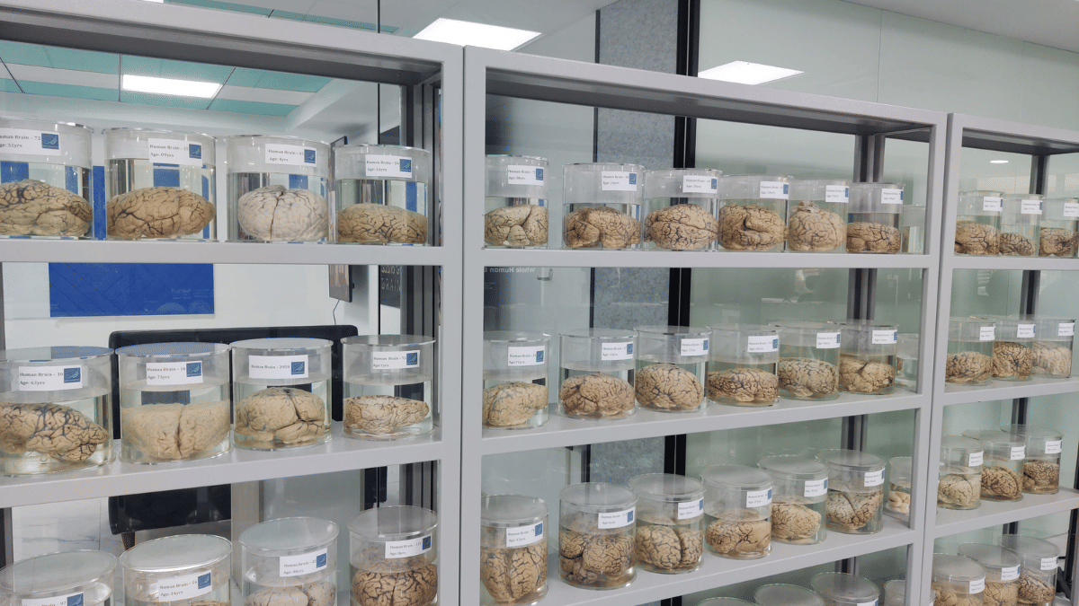
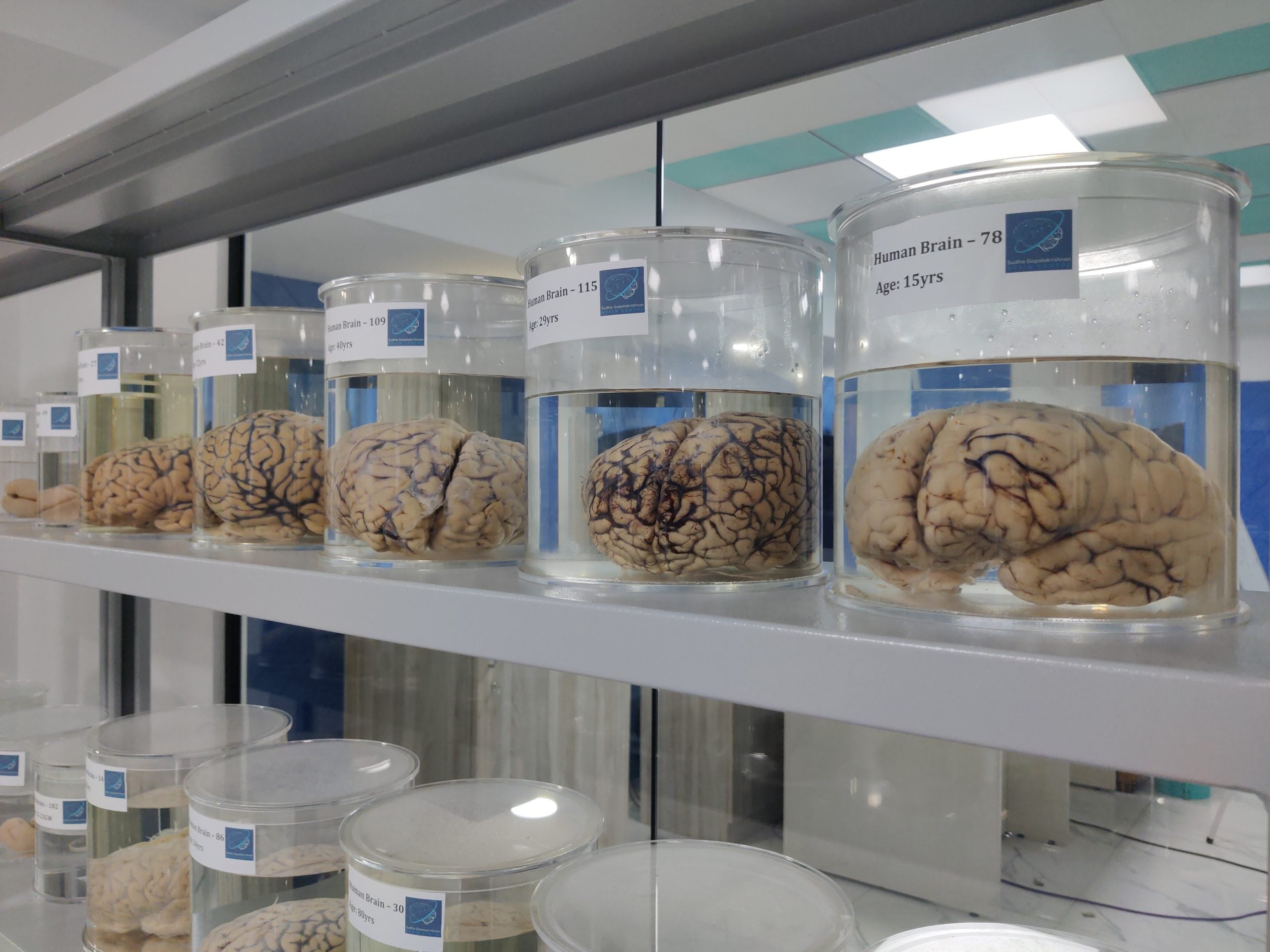
Chennai: Two grey metal shelves stand against a glass partition in the lobby of the Sudha Gopalakrishnan Brain Centre (SGBC) at IIT Madras. Clear acrylic jars filled with fluid hold human brain specimens in neat rows.
Each jar has a white label with the centre’s logo and a number, along with details such as “Human Brain–58, Age: 18 yrs” and “Human Brain–33, Age: 60 yrs”. The adult brains are pale brown, with fine bluish veins visible across their folded surfaces. At one end of each shelf are smaller jars labelled “Fetal Brain”. Inside one, a fetal brain about the size of a plum has a smooth surface, without the deep folds seen in adult brains.
Along a white marble corridor, a navy-blue wall titled “Whole Human Brain Histopathology Pipeline” shows the steps involved in preparing a brain for study, from removing and freezing the tissue to cutting it into thin slices, staining it and capturing images under a microscope.
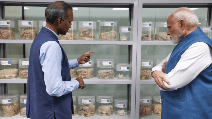
The jars are the starting point of a project whose real scale becomes visible on computer screens. On 11 October 2026, Prime Minister Narendra Modi launched SGBC’s mission to map more than 100 human brains into three-dimensional atlases at the level of individual cells. The project covers different stages of life, from fetal development to old age, and includes brains affected by diseases.
The SGBC also released C-STROKE, described as the world’s first three-dimensional atlas of a human brain affected by an ischaemic stroke, which occurs when a blockage cuts off blood supply to part of the brain.
According to Prof Mohanasankar Sivaprakasam, head of SGBC, the whole-brain, 3D, cell-resolution data available across the world is limited to around 20 or 30 brains at most.
“We are the first people to attempt such a large-scale mapping across human life,” he told ThePrint. “That is happening because of the technology platform we have developed, which allows you to do it at such a scale.”
Unlike the heart, whose functioning is relatively well understood, the brain is harder to study because it is enclosed within the skull. Doctors can examine it using magnetic resonance imaging (MRI) or measure its electrical activity through EEG, but these methods are limited to millimeters and centimeters. They cannot reveal its workings down to individual cells.
MRI captures structures at a scale of about a millimetre, while a single neuron is roughly 10 microns across, about 100 times smaller. This gap in resolution limits how much doctors and researchers can see of the brain’s structure and functioning, leaving important questions in neurology and neurosurgery unanswered, Mohanasankar said.
Also read: How IIT Madras is mapping India’s ‘vascular age’ to develop early warning guidelines for heart risks
‘A technology gap’
The seeds of this process were sown around 2015, Mohanasankar explained, when Infosys co-founder and IIT Madras alumnus Kris Gopalakrishnan began exploring ways to support brain research after stepping away from the company. He established three chair professorships at the institute.
By 2017-18, the discussions had moved towards a more ambitious question of whether researchers can map an entire human brain, cell by cell. Gopalakrishnan wanted the project to combine neuroscience, the study of the brain and nervous system, with engineering, drawing on India’s engineering expertise.
“We have been doing good science in the field of neuroscience, but we haven’t taken up very large, challenging mission-type projects in the country,” he said.
The team examined research being done around the world. Scientists were mapping mouse brains, while studies of monkey and human brains often focused on particular regions rather than the entire organ.
“We asked, why are people not doing human brain mapping? The whole brain,” Mohanasankar said. “We realised it is actually a technology gap and an engineering challenge.”
The difficulty lies partly in the difference between what a hospital scan can show and what researchers need to see. MRI produces images at a scale of a few millimetres. Individual brain cells are much smaller.
To study individual cells across an entire brain, researchers must acquire brains collected after death. These brains are acquired at collaborating medical institutions through a specialised process developed by the team.
They freeze the brain, cut it into extremely thin slices, use special chemicals to make different cells and structures visible, and photograph each slice under specially designed large scale microscopes. The images are then combined digitally to reconstruct the brain in three dimensions.
But cutting the brain is one of the most difficult parts of the process.
An adult human brain must be cut into around 10,000 slices, each about 10-20 microns thick. To put that thickness in perspective, a strand of human hair is several times thicker. The brain slices are roughly half the size of a sheet of paper, yet are so thin that they can easily tear or break apart during handling.
Damaged slices can leave gaps in the final reconstruction. If even a few percent of the slices are lost, it can compromise the map of the critical regions, especially in diseased brains. Researchers must therefore develop ways to freeze the tissue, cut it and transfer each delicate slice onto a glass slide without damaging it.
Even intact slices can change shape slightly around the edges. This makes it harder for computers to line up the images correctly and rebuild the brain at the scale of Gigapixels and Teravoxels.
Mohanasankar explained that the team had to develop specialised equipment and software to handle the tissue and correct these distortions. Mapping a single adult brain can take four to eight months, depending on its size and the time needed to process the tissue and images.
The process also generates enormous amounts of information. A single brain can produce several petabytes of data, Mohanasankar said. One petabyte is roughly equivalent to one million gigabytes. Storing, processing and searching through so much information requires substantial computing power.
SGBC has built an imaging and computing system to turn whole human brains into digital maps at cellular resolution. The SGBC has also developed tools that allow researchers to explore these petabyte scale maps on computers and mobile phones, rather than requiring them to work directly with the original tissue or operate a supercomputer.
“Its AI-driven computing and imaging pipeline enables petabyte-scale datasets to be analysed within hours rather than days,” Mohansankar explained.
In 2019, Gopalakrishnan agreed to support the project despite the risks. “It’s extremely risky, but I will support it,” Mohanasankar recalled him saying.
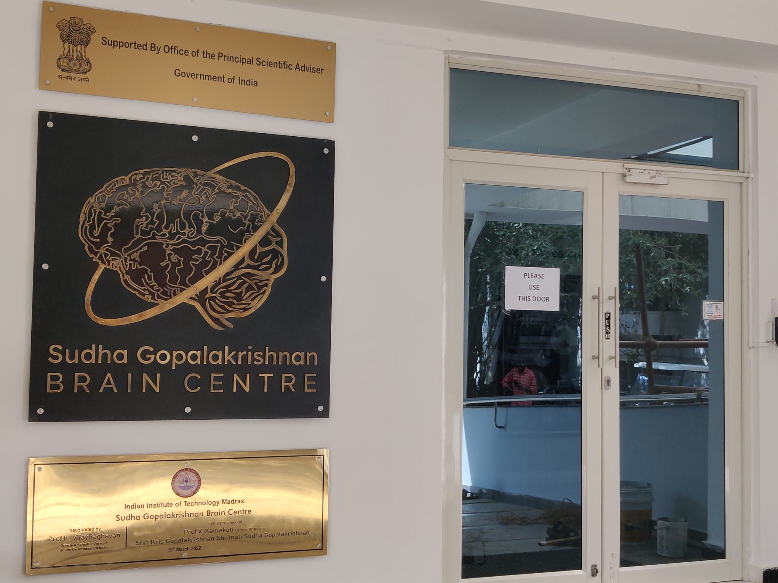
The proposal was submitted to the Office of the Principal Scientific Adviser, then headed by K. Vijayraghavan, and approved in February 2020. Within weeks, the Covid-19 pandemic disrupted work.
The team began in March 2020 without a laboratory and amid restrictions on movement. Much of the technology platform was developed during the pandemic, Mohanasankar said.
From Dharani to C-STROKE
The centre’s first atlas, Dharani, was released in December 2024-January 2025. It maps the developing brain during the second trimester of pregnancy, covering approximately 12-24 weeks of gestation, when the brain grows rapidly and its structures develop.
An editorial in the Journal of Comparative Neurology described the work with the headline “India gets a seat at the table of human brain cartography”.
In June 2026, SGBC released ‘Anchor’, an atlas of the brainstem, the part of the brain that connects to the spinal cord and helps regulate essential functions such as breathing and heart rate. The atlas includes fetal, child and adult brains. The brainstem contains densely packed nerve cells and other structures, making it particularly difficult to map in detail, Mohanasankar said.
C-STROKE applies the same approach to understanding stroke. Doctors can use MRI scans to identify where a stroke has occurred, but the scans cannot show all the changes taking place inside individual cells.
The atlas links MRI scans taken before death with detailed images of brain tissue examined afterwards. This allows researchers to compare what a scan shows with the damage visible inside the tissue.
It contains more than 1,000 labelled sections from a brain affected by an ischaemic stroke. These sections map signs of damage to nerve cells, inflammation, disruption of the blood-brain barrier, low oxygen levels and injury to nerve fibres. The blood-brain barrier is a protective layer that controls which substances can pass from the bloodstream into the brain.
“By examining these changes, researchers hope to understand what the abnormalities seen on clinical scans mean at the cellular level. This could help them investigate how stroke damages the brain and identify possible targets for treatment,” Mohanasankar said.
The three-dimensional maps were generated using the Param Shakti supercomputer, with support from the Ministry of Electronics and Information Technology. The research is supported by Prem Watsa, an IIT Madras alumnus and chairman of Fairfax Financial.
An online viewer and analytical tools allow researchers and clinicians to explore the atlas and study the tissue at different levels of detail.
The larger goal is to understand how diseases affect the brain and use that knowledge to guide research into treatments, including drugs and gene therapies. More detailed maps could also help researchers study how damage affects particular brain structures and potentially improve planning for certain surgeries.
SGBC has collected more than 300 brains, from institutions CMC Vellore, KMC, SMCH, Mediscan in Chennai, NIMHANS and centres in Gujarat. The collection includes brains affected by Alzheimer’s disease, rabies and other forms of neurodegeneration, as well as congenital conditions such as holoprosencephaly. In this condition, the developing brain does not divide fully into two halves.
Comparing brains without known disease with those affected by different conditions could help researchers identify patterns that are difficult to detect through conventional scans or by examining small sections of tissue alone.
“We expect more findings to emerge over the next one to two years as the team uses computational methods to analyse the growing collection,” Mohanasankar told ThePrint.
The people behind the maps
Asked what part of the work had received too little attention, Mohanasankar pointed away from the technology.
“How does such a work get done in India?” he said.
The project depends on doctors and medical institutions that help collect and document brain tissue. Fetal medicine specialists and obstetricians document fetal cases, many involving deaths before birth. Forensic doctors help collect adult brain tissue, while neurologists and neurosurgeons contribute their medical expertise.
“Many of these doctors are so busy… for them to do this selflessly, that is really something,” he said.
Families which agree to donate brain tissue are another important part of the effort. Some donations have come from families of young adults with multiple sclerosis, a disease in which the immune system damages the protective covering around nerve fibres.
“For them to come forward and donate these brains… so many people have come together,” he said.
The mission brings together more than 200 researchers, engineers and technicians at SGBC, over 10 medical institutions across India and more than 30 researchers from over 10 countries.
“This is perhaps the first time such a large-scale global science project in India has received CSR and philanthropy funding from over 20 organisations,” Mohanasankar said.
This reporter visited IIT Madras on an invitation from the institution.
(Edited by Viny Mishra)