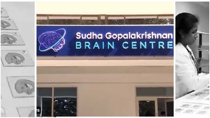
Chennai: In an ambitious new initiative, the Indian Institute of Technology (IIT) Madras has launched a brain research centre that aims to build detailed cellular-level 3D maps of the human brain to better understand neural connections as well as how some diseases affect the brain.
While MRI scans allow scientists to peek into a living brain, at the cellular level there is still a lot left to learn.
Ahead of its launch Friday, the new Sudha Gopalakrishnan Brain Centre, led by IIT Madras professor Mohanasankar Sivaprakasam, already completed the imaging process of three developing human brains.
The Centre is supported by tech billionaire Kris Gopalakrishnan and his spouse Sudha Gopalakrishnan. However, the size of the funding provided was not disclosed by the duo or the IIT-M authorities.
Also read: ‘Where’s the research?’ Why space institute IIST’s tailor-made graduates don’t stay at ISRO
How the imaging will work
The entire brain imaging process is divided into a series of steps. Equipment to carry out each of the steps was either built from scratch at IIT Madras, or are off the shelf products that have been modified at the institute, IIT-M Director V. Kamakoti told ThePrint.
Firstly, the IIT-M team worked with several institutes, including Christian Medical College, Vellore, and National Institute of Mental Health and Neurosciences (NIMHANS), Bengaluru, to acquire human brains.
The first challenge was to acquire human brains, Sivaprakasam told ThePrint. “Not only does it have to be extracted within the first few hours of death, it also has to be extracted without any damage,” he said.
Soon after this the whole brain needs to be frozen. To do this, the team did a 3D-scan of the entire brain and printed a metallic replica. This replica is then used to create a mould in which the brain can fit perfectly.
The brain is then dipped into a cryogenic fluid of -80 degrees, which instantly freezes the brain.
The next challenge is to slice the brain in slices just 20 microns thick, and transfer these onto slides that can be imaged under the microscope.
For this, the brain is placed in a modified cryogenic tank at -20 degrees, where a special adhesive tape is used to pull out thin slices of the brain. The adhesive sheet is then placed on a glass slide, and treated with UV light, which causes the thin brain slice to get deposited on the glass slide.
Since the human brain is rarely sliced and studied in this manner, the team had to work with glass slides big enough to mount a cross-section of the human brain which are much larger than usual slides. This came with an additional challenge of covering the slide with what is known as a cover slip, which essentially preserves the sample from damage.
Each brain when sliced will lead to over 7,000 slides. Preparing these slides by hand is not only a slow and tedious process, but can also lead to air bubbles getting trapped in the slides, which could interfere with the imaging process. To overcome this problem, the researchers designed a system that automates the process of placing the cover slip on the slide.
The images of slides obtained from one brain can take up over hundreds of terabytes of space.
Eventually, the researchers ‘stitch together’ the individual brain slice images to create a complete map of the human brain. This is where big data analytics come in, allowing scientists to understand the connections in the brain at the cellular level and over time understand how different diseases affect the brain.
Sivaprakasam said that his team is in touch with medical institutions to acquire more post-mortem human brains. His aim is to be able to image 10 brains over the next two years.
What the PSA said
Addressing the press at the inaugural event Friday, K. VijayRaghavan, Principal Scientific Adviser to the Government of India, said the IIT Madras brain centre will help in solving complex issues that will benefit the world.
“The combination of IIT Madras, which has the expertise in science and data analysis, with medicine is going to be revolutionary. Going forward, we have an extraordinary problem in Neuroscience, i.e. on the functioning of human brain. We are at an earlier stage in our understanding of the human brain functioning,” he said.
“The dynamic leadership of IIT Madras has shown the ability to herd different kinds of complex talent together. The IIT Madras Research Park is an example and today every institution wants to copy the model,” he added.
The PSA’s office will support the project — titled ‘Computational and Experimental Platform for High-Resolution Terapixel Imaging of ex-vivo Human Brains’ — for high-throughput light microscopic imaging of whole human brains till the team is able to develop a proof of concept required to apply for further funding.
George Varghese, a CMC Vellore professor who is collaborating with IIT-M on the project, told ThePrint that the project will help understand how diseases like rabies affects the brain — knowledge that may help pave the way for new treatments.
Also read: From body to brains and personality, here’s how humans will look like after 10,000 years


